lv diverticulum | left ventricular diverticulum lv diverticulum A left ventricular diverticulum is an infrequent heart condition that affects the muscle wall of the left ventricle. It is brought on by a bag-like structure or pouch-like . Yes, most LV handbags are usually made from real animal skin. Other bags that are made of other materials usually feature leather trims and handles. Python and other exotic animal skins are also used. Some handbags are also made from ostrich skins, stingray skins, and crocodile skins.
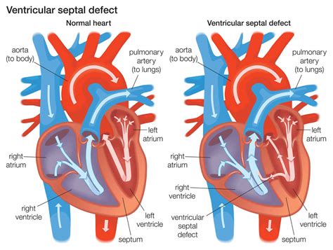
0 · vadcume despamed lvd
1 · septum ventricular
2 · myocardial diverticulum
3 · lv diverticulum vs aneurysm
4 · lv apical diverticulum
5 · left ventricular diverticulum echo
6 · left ventricular diverticulum
7 · inferoapical wall
My Visit. Save time by preparing for your appointment ahead of time. Complete patient forms, check our list of accepted insurances, and get answers to all of your questions here.
Congenital cardiac diverticulum is an outpouching of the three cardiac layers, the endocardium, myocardium, and pericardium. A rare presentation from the left ventricle, this is usually found in infancy with rare presentation in adulthood. Risk factors include genetic . A left ventricular diverticulum is an infrequent heart condition that affects the muscle wall of the left ventricle. It is brought on by a bag-like structure or pouch-like . Cardiac diverticula most commonly occur in the left ventricle but have been reported to occur in all chambers of the heart. Despite reports of their rare occurrence, cardiac . This congenital left ventricular apical diverticulum was found by chance by means of an echocardiographic study to elucidate the etiology of systolic murmur, and its diagnosis .
Congenital left ventricular diverticulum (LVD) is a rare cardiac malformation. Its prevalence rate is less than 0.1% of the congenital heart diseases requiring surgery. Some scholars suggest that .We aim to discuss the literature related to the left ventricular diverticulum, its presentation and the treatment options. The pathogenesis of ventricular diverticulum remains unknown, although .
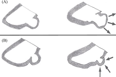
LV diverticula are classified as apical or nonapical, on the basis of their location: apical diverticula are more prevalent and described as finger-like or hook-like contractile .True congenital diverticulum of the left ventricle (LV) is seen very rarely in adults. We report a case of congenital LV diverticulum in an adult patient, in whom clinical findings and imaging .We present two cases of LV diverticulum in adult patients that illustrate the characteristic features of diverticula and highlight the advantages of using echocardiographic contrast combined with 2-dimensional harmonic imaging . Congenital cardiac diverticulum is an outpouching of the three cardiac layers, the endocardium, myocardium, and pericardium. A rare presentation from the left ventricle, this is usually found in infancy with rare presentation in adulthood. Risk factors include genetic history and being male.
A left ventricular diverticulum is a pouch or sac branching out from the ventricle. They have a variable size and can range from 5 mm to 80-90 mm. It is thought to arise as a developmental anomaly, from around the 4 th embryonic week. A left ventricular diverticulum is an infrequent heart condition that affects the muscle wall of the left ventricle. It is brought on by a bag-like structure or pouch-like protuberance, including the muscle wall's endocardium, myocardium, and pericardium. Cardiac diverticula most commonly occur in the left ventricle but have been reported to occur in all chambers of the heart. Despite reports of their rare occurrence, cardiac ventricular diverticula are fairly common findings in patients undergoing cardiac . This congenital left ventricular apical diverticulum was found by chance by means of an echocardiographic study to elucidate the etiology of systolic murmur, and its diagnosis was established by enhanced computed tomography.
Congenital left ventricular diverticulum (LVD) is a rare cardiac malformation. Its prevalence rate is less than 0.1% of the congenital heart diseases requiring surgery. Some scholars suggest that all LVD should be actively removed to prevent .
vadcume despamed lvd

septum ventricular
We aim to discuss the literature related to the left ventricular diverticulum, its presentation and the treatment options. The pathogenesis of ventricular diverticulum remains unknown, although most authors consider it to be a malformation. LV diverticula are classified as apical or nonapical, on the basis of their location: apical diverticula are more prevalent and described as finger-like or hook-like contractile pouches, typically less than 3 cm in length and 1.25 cm in width, .

True congenital diverticulum of the left ventricle (LV) is seen very rarely in adults. We report a case of congenital LV diverticulum in an adult patient, in whom clinical findings and imaging studies suggested a postinfarction pseudoaneurysm.
We present two cases of LV diverticulum in adult patients that illustrate the characteristic features of diverticula and highlight the advantages of using echocardiographic contrast combined with 2-dimensional harmonic imaging when considering the differential diagnosis of this condition. Congenital cardiac diverticulum is an outpouching of the three cardiac layers, the endocardium, myocardium, and pericardium. A rare presentation from the left ventricle, this is usually found in infancy with rare presentation in adulthood. Risk factors include genetic history and being male. A left ventricular diverticulum is a pouch or sac branching out from the ventricle. They have a variable size and can range from 5 mm to 80-90 mm. It is thought to arise as a developmental anomaly, from around the 4 th embryonic week.
A left ventricular diverticulum is an infrequent heart condition that affects the muscle wall of the left ventricle. It is brought on by a bag-like structure or pouch-like protuberance, including the muscle wall's endocardium, myocardium, and pericardium. Cardiac diverticula most commonly occur in the left ventricle but have been reported to occur in all chambers of the heart. Despite reports of their rare occurrence, cardiac ventricular diverticula are fairly common findings in patients undergoing cardiac . This congenital left ventricular apical diverticulum was found by chance by means of an echocardiographic study to elucidate the etiology of systolic murmur, and its diagnosis was established by enhanced computed tomography.
Congenital left ventricular diverticulum (LVD) is a rare cardiac malformation. Its prevalence rate is less than 0.1% of the congenital heart diseases requiring surgery. Some scholars suggest that all LVD should be actively removed to prevent .We aim to discuss the literature related to the left ventricular diverticulum, its presentation and the treatment options. The pathogenesis of ventricular diverticulum remains unknown, although most authors consider it to be a malformation. LV diverticula are classified as apical or nonapical, on the basis of their location: apical diverticula are more prevalent and described as finger-like or hook-like contractile pouches, typically less than 3 cm in length and 1.25 cm in width, .
True congenital diverticulum of the left ventricle (LV) is seen very rarely in adults. We report a case of congenital LV diverticulum in an adult patient, in whom clinical findings and imaging studies suggested a postinfarction pseudoaneurysm.

myocardial diverticulum

pink rhinestone gucci sunglasses replica
For each heads, remove an Energy card attached to the Defending Pokémon and put it in the Lost Zone . Put this card onto your Active Dialga . Dialga LV.X can use any attack, Poké-Power, or Poké-Body from its previous Level.
lv diverticulum|left ventricular diverticulum


